JY/T 0587-2020 PDF EnglishUS$485.00 · In stock · Download in 9 seconds
JY/T 0587-2020: General rules for X-ray polycrystalline diffractometry Delivery: 9 seconds. True-PDF full-copy in English & invoice will be downloaded + auto-delivered via email. See step-by-step procedure Status: Valid
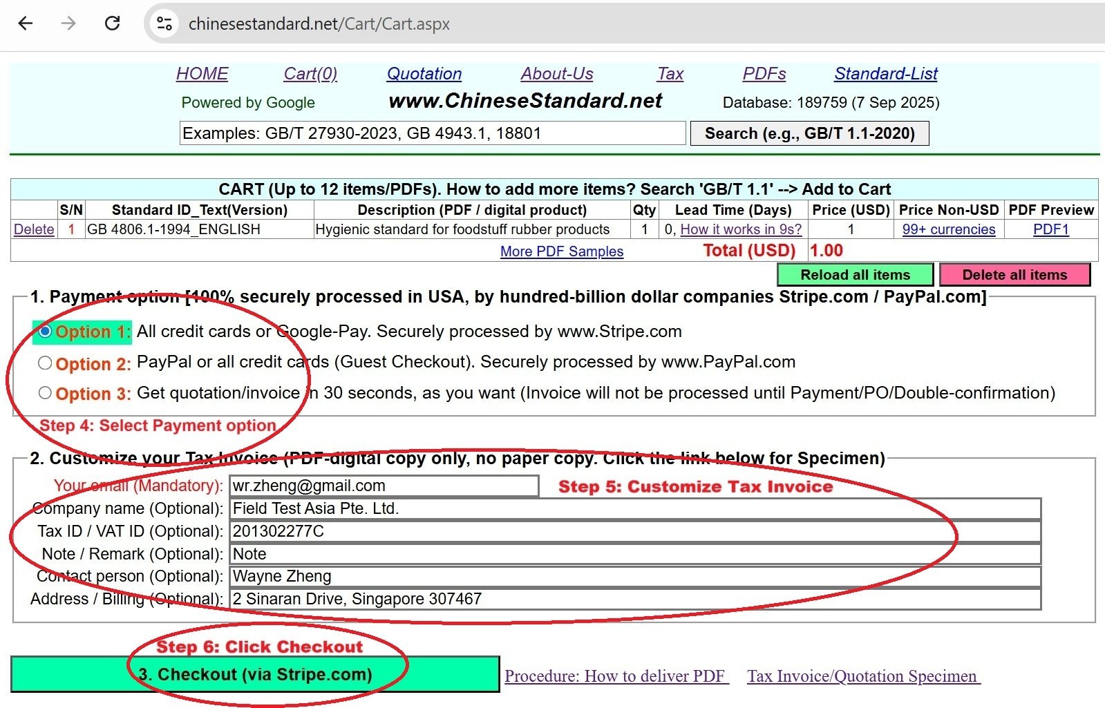
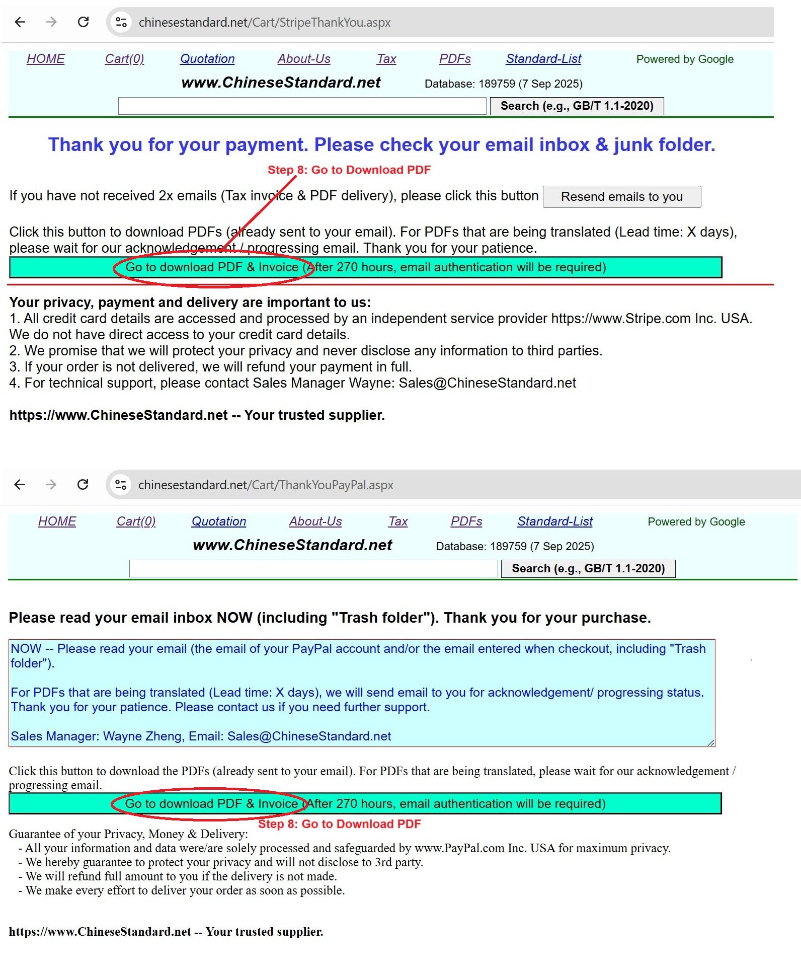
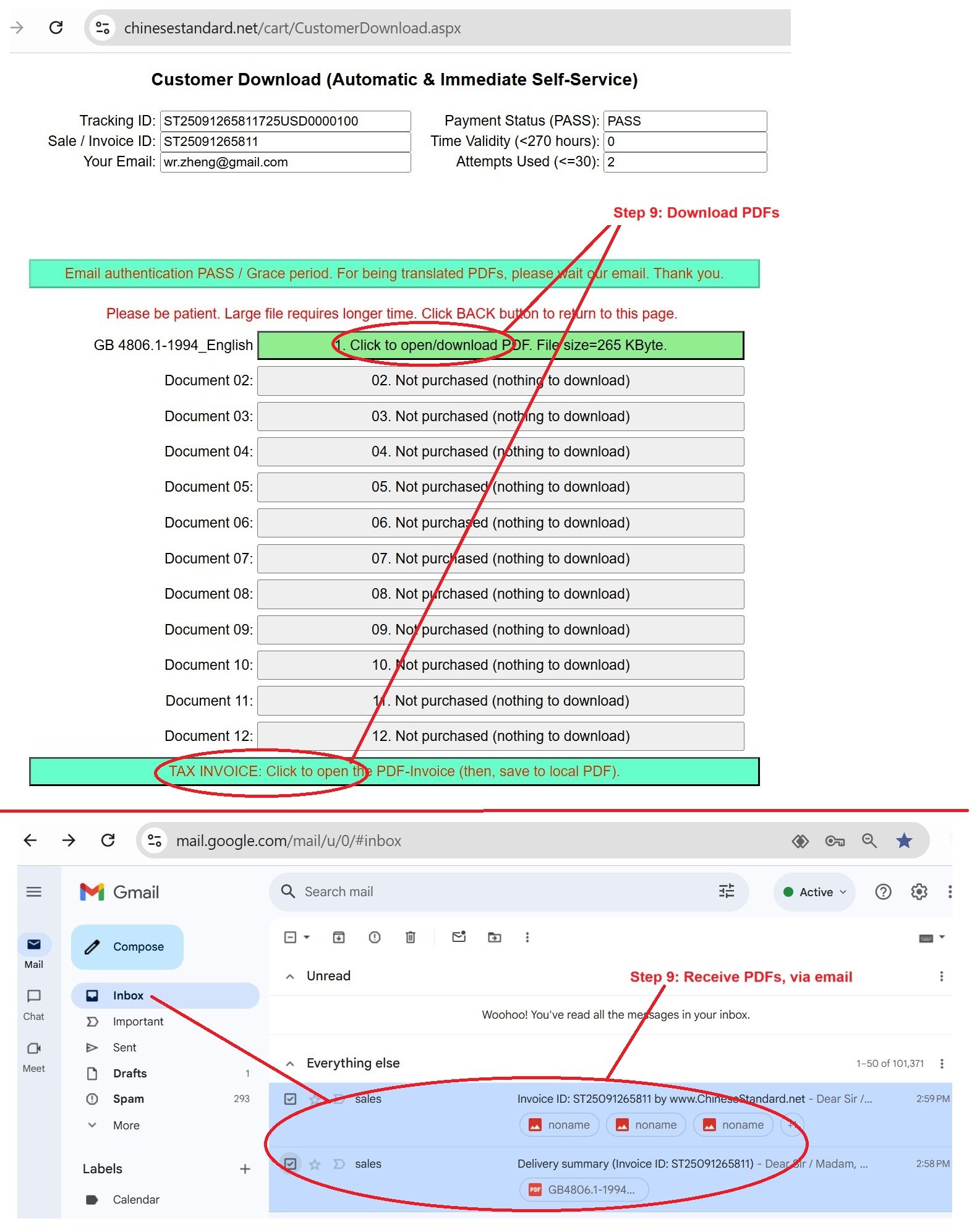
JY/T 0587-2020: General rules for X-ray polycrystalline diffractometry---This is an excerpt. Full copy of true-PDF in English version (including equations, symbols, images, flow-chart, tables, and figures etc.), auto-downloaded/delivered in 9 seconds, can be purchased online: https://www.ChineseStandard.net/PDF.aspx/JYT0587-2020EDUCATION INDUSTRY STANDARD ICS 03.180 Y 51 Replacing JY/T 009-1996 General rules for X-ray polycrystalline diffractometry Issued on. SEPTEMBER 29, 2020 Implemented on. DECEMBER 01, 2020 Issued by. Ministry of Education of PRC Table of ContentsForeword... 4 1 Scope... 6 2 Normative references... 6 3 Terms and definitions... 6 4 Principles of analysis methods... 11 4.1 Qualitative analysis of phases... 11 4.2 Quantitative analysis of phase... 12 4.3 Determination of grain size and lattice distortion... 13 4.4 Determination of unit cell parameters of cubic crystals... 15 4.5 Solving the crystal structure from ab initio polycrystalline diffraction data... 16 4.6 High and low temperature diffraction... 16 5 Reagents and materials... 17 5.1 Standard material... 17 5.2 Organic solvents... 17 5.3 Sieves... 17 5.4 Sample preparation tools... 17 5.5 Microscope... 17 5.6 Specimen plate... 17 6 Instruments... 18 6.1 Composition of the instrument... 18 6.2 Verification or calibration... 18 7 Samples... 19 7.1 Pretreatment of samples... 19 7.2 Filling of the specimen plate... 19 7.3 Judge whether the specimen plate is available... 20 8 Analysis steps... 20 8.1 Startup and parameter setting of the instrument... 20 8.2 Steps of phase qualitative analysis... 21 8.3 Steps for phase quantitative analysis... 23 8.4 Determination of grain size and lattice distortion by linewidth method... 24 8.5 Procedure for determination of unit cell parameters of cubic crystals... 25 8.6 Analysis steps for solving crystal structure by ab initio polycrystalline diffraction data ... 26 8.7 High temperature and low temperature polycrystalline diffraction... 27 8.8 Crystallinity analysis procedure... 28 8.9 Inspection after determination... 29 9 Results report... 29 9.1 Basic information... 29 9.2 Presentation of analysis results... 29 10 Safety precautions... 30 10.1 X-ray protection... 30 10.2 Water and electricity safety protection... 30 10.3 Safety protection for experiment personal... 30 Appendix A (Informative) PDF descriptions... 32 Appendix B (Informative) Various standard materials and standard data... 36 Appendix C (Informative) Calibration of diffraction peak positions (2θ)... 40 Appendix D (Informative) Crystallinity analysis method... 44 References... 481 ScopeThis standard specifies the analysis method principles, reagents and materials, instruments, samples, analysis steps, result reports, safety precautions, for using polycrystalline X-ray diffractometers, to analyze the phase composition of various polycrystalline materials. This standard applies to conventional polycrystal X-ray diffractometers. X-ray diffractometers, which are equipped with two-dimensional surface detectors, can refer to this method.2 Normative referencesThe following documents are essential for the application of this document. For dated references, only the dated version applies to this document. For undated references, the latest edition (including all amendments) applies to this document. GB/T 13869-2008 General guide for safety of electric user GB 18871-2002 Basic standards for protection against ionizing radiation and for the safety of radiation sources JY/T 009-1996 General rules for X-ray polycrystalline diffractometry3 Terms and definitionsThe terms and definitions, which are defined in JY/T 009-1996, as well as the following terms and definitions, apply to this document. 3.1 X-ray Electromagnetic waves, which have wavelengths from 10-3 nm to 10 nm. Note. The X-ray wavelength, which is used for crystal diffraction, is 0.05 nm ~ 0.25 nm. 3.2 Crystal A generalized crystal is a solid, which has a definite diffraction pattern. Its atoms, molecules or ions are arranged in the space a highly ordered manner, according to certain rules, including traditional periodic crystals and non-periodic crystals. 3.3 Polycrystal A solid powder or bulk object, which is formed by agglomeration of many small grains. It is also known as polycrystalline material. 3.4 Space lattice In crystallography, the tool used to express the periodic arrangement of structural units in a crystal, which is a collection of periodically repeating points in three- dimensional space. 3.5 Unit cell The smallest building unit, in which atoms, molecules or ions are regularly arranged, in long-range order, in three-dimensional space. Its shape is a parallelepiped.4 Principles of analysis methods4.1 Qualitative analysis of phases The process, according to the X-ray diffraction pattern (peak position, intensity, element composition) of the sample to be analyzed, to search and match with the powder diffraction database, to compare and analyze to determine its phase composition. 4.2 Quantitative analysis of phase 4.2.1 Basic formulas for quantitative phase analysis The diffraction spectrum of the polycrystalline mixture of different phases is the superposition of the weight (scale factor) of the diffraction spectrum of each constituent phase. The diffraction intensity (referring to the full spectrum intensity) of each constituent phase is affected by other phases (referring to the total mass absorption coefficient), 4.3.2 Relationship between lattice distortion or micro-strain and diffraction line width 4.4 Determination of unit cell parameters of cubic crystals 4.4.1 The relationship between the unit cell parameters of the cubic material and the Bragg angle θhkl of a certain hkl diffraction The relationship, between the unit cell parameters of the cubic material and the Bragg angle θhkl of a certain hkl diffraction, is.5 Reagents and materials5.1 Standard material The most commonly used is silicon powder; the purity is better than 99.999%; the particle size is between 5 μm and 30 μm; the crystal is perfect; there is no residual stress and too many defects. Silica powder is suitable for the range of 2θ > 29°. 5.4 Sample preparation tools Microscope slides, flat glass, agate mortars, clips and other sample preparation supplies. 5.5 Microscope An optical microscope, which is used to observe the uniform particle size of the sample AND determine whether the specimen plate is flat. 5.6 Specimen plate Specimen plates without backing, such as hollow aluminum specimen plate, grooved glass specimen plate, single crystal silica, or porous material.6 Instruments6.1 Composition of the instrument 6.1.1 Structure diagram Polycrystalline X-ray diffractometer mainly consists of four parts. X-ray generator, Bragg-Brentano goniometer (vertical or horizontal), detection and recording system, control and data processing system. Its structure diagram is as shown in Figure 1. 6.2 Verification or calibration Before the equipment is put into use, it shall verify and calibrate, in a planned manner, the equipment that has a significant impact on the accuracy or validity of the detection and analysis results, including auxiliary measurement equipment used to measure environmental conditions, etc., to determine whether it meets the requirements of detection and analysis.7 Samples7.1 Pretreatment of samples The pretreatment of the sample includes grinding, polishing, etc. Equipment for grinding samples includes pulverizers, ball mills, mortars, etc. Different grinding equipment and accessories are selected, according to the hardness of the analyzed specimen AND the influence of impurities that may be introduced. 7.1.1 Grinding 7.1.3 Judging whether the mixture is uniform Take specimens, which have different mixing times, to perform diffraction scanning. If the obtained spectrum is basically the same, it means that the mixing is uniform. If the diffraction intensity of different phases, on the obtained spectrum, changes greatly, it means that the mixing is not uniform, AND it is necessary to continue mixing. 7.2 Filling of the specimen plate 7.2.1 Usually, use a grooved specimen plate. Evenly fill the specimen in the groove. The surface of the specimen is slightly higher than the surface of the specimen plate. Use a glass slide to press the specimen tightly, so that the surface of the specimen and the surface of the specimen plate are on the same plane. 7.3 Judge whether the specimen plate is available Quickly scan the specimen plate. If the diffraction line intensity sequence of each substance is similar to that listed in the PDF card, it means that there is no serious preferred orientation, so this specimen plate is available. Otherwise, the specimen plate shall be refilled.8 Analysis steps8.1 Startup and parameter setting of the instrument 8.1.1 Power on Start the instrument, according to the instrument operating procedures. 8.1.2 Setting of instrument parameters 8.1.2.4 Scan mode and scan speed There are two common scanning methods. continue scanning and step scanning. Scanning speed refers to the angular speed, at which the receiving slit (RS) and the detector rotate uniformly, on the goniometer, in °/min. Increasing the scanning speed can save test time, BUT will result in a decrease in intensity and resolution. 8.2 Steps of phase qualitative analysis 8.2.1 Preparation before measurement 8.2.1.1 Power on and instrument calibration Start the instrument, according to the provisions of 8.1.Set the parameters. Determine whether the instrument is verified or calibrated, according to the provisions of 6.2. 8.2.1.2 Specimen preparation Prepare specimens, according to the relevant provisions in Chapter 7. 8.2.2 Determination 8.2.4 Spectrum analysis 8.2.4.1 Retrieving the phase contained in the matching specimen Use PDF or CCDC index for automatic computer retrieval or manual retrieval, to find out possible diffraction cards or other patterns of known phases. Make careful comparison; finally determine the phase contained in the specimen. 8.2.4.2 Reducing influencing factors During the analysis, attention shall be paid to the large shift of the d value or the relative intensity data, due to the influence of the solid solution phenomenon, the overlapping peaks of the mixture, the preferred orientation, etc. If there is an obvious preferred orientation, consideration shall be given to re-preparing the specimen OR using a rotating specimen stage, during the measurement, to reduce its influence. 8.2.4.5 Analyzing trace phases in the specimen If the specimen contains trace phase, it is best to obtain the trace phase single-phase sample by extraction and enrichment method; conduct phase analysis on it. If extraction is impossible, it may use the methods, such as increasing the radiation power or extending the data collection time, to make the specimen appear as 3 or more diffraction lines as possible, to carry out the phase analysis. 8.3 Steps for phase quantitative analysis 8.3.1 Preparation before determination 8.3.3.2 Modeling Transfer the diffraction spectrum into the Rietveld full spectrum fitting software. Input the crystal structure model, peak shape model, texture model, background model of the diffraction spectrum, of each constituent phase. 8.3.3.3 Refinement Rietveld full spectrum fitting refinement. 8.4 Determination of grain size and lattice distortion by linewidth method 8.4.1 Preparation before determination 8.4.2 Determination Scan the sample for testing. If the intensity of the diffraction peak is lower than 1 x 10⁴ counts, THEN, the dwell time in each step shall be appropriately extended. 8.4.3 Spectral processing 8.4.3.1 Selection of diffraction peaks and calculation of full width at half maximum of peak profile Test the linewidth B of each diffraction line of the standard sample AND each diffraction line of the sample to be tested. 8.4.3.2 Calculation of βa and βd By separating the linewidths of diffraction lines and removing the influence of instrument broadening, the grain broadening βa and lattice distortion broadening βd are obtained, respectively. the operating procedures of the instrument.9 Results report9.1 Basic information For any determinations, which are specified in this standard, the result report shall at least include. information on the entrusting organization, sample information, equipment information, environmental conditions, testing method (standard), testing person, testing date, etc. 9.2 Presentation of analysis results List the results obtained and the relative errors. For the analysis results given, it shall describe the measurement method used and the actual treatment process. List the standard samples used and their proportions. JY/T 0587-2020 EDUCATION INDUSTRY STANDARD ICS 03.180 Y 51 Replacing JY/T 009-1996 General rules for X-ray polycrystalline diffractometry Issued on. SEPTEMBER 29, 2020 Implemented on. DECEMBER 01, 2020 Issued by. Ministry of Education of PRCTable of ContentsForeword... 4 1 Scope... 6 2 Normative references... 6 3 Terms and definitions... 6 4 Principles of analysis methods... 11 4.1 Qualitative analysis of phases... 11 4.2 Quantitative analysis of phase... 12 4.3 Determination of grain size and lattice distortion... 13 4.4 Determination of unit cell parameters of cubic crystals... 15 4.5 Solving the crystal structure from ab initio polycrystalline diffraction data... 16 4.6 High and low temperature diffraction... 16 5 Reagents and materials... 17 5.1 Standard material... 17 5.2 Organic solvents... 17 5.3 Sieves... 17 5.4 Sample preparation tools... 17 5.5 Microscope... 17 5.6 Specimen plate... 17 6 Instruments... 18 6.1 Composition of the instrument... 18 6.2 Verification or calibration... 18 7 Samples... 19 7.1 Pretreatment of samples... 19 7.2 Filling of the specimen plate... 19 7.3 Judge whether the specimen plate is available... 20 8 Analysis steps... 20 8.1 Startup and parameter setting of the instrument... 20 8.2 Steps of phase qualitative analysis... 21 8.3 Steps for phase quantitative analysis... 23 8.4 Determination of grain size and lattice distortion by linewidth method... 24 8.5 Procedure for determination of unit cell parameters of cubic crystals... 25 8.6 Analysis steps for solving crystal structure by ab initio polycrystalline diffraction data ... 26 8.7 High temperature and low temperature polycrystalline diffraction... 27 8.8 Crystallinity analysis procedure... 28 8.9 Inspection after determination... 29 9 Results report... 29 9.1 Basic information... 29 9.2 Presentation of analysis results... 29 10 Safety precautions... 30 10.1 X-ray protection... 30 10.2 Water and electricity safety protection... 30 10.3 Safety protection for experiment personal... 30 Appendix A (Informative) PDF descriptions... 32 Appendix B (Informative) Various standard materials and standard data... 36 Appendix C (Informative) Calibration of diffraction peak positions (2θ)... 40 Appendix D (Informative) Crystallinity analysis method... 44 References... 481 ScopeThis standard specifies the analysis method principles, reagents and materials, instruments, samples, analysis steps, result reports, safety precautions, for using polycrystalline X-ray diffractometers, to analyze the phase composition of various polycrystalline materials. This standard applies to conventional polycrystal X-ray diffractometers. X-ray diffractometers, which are equipped with two-dimensional surface detectors, can refer to this method.2 Normative referencesThe following documents are essential for the application of this document. For dated references, only the dated version applies to this document. For undated references, the latest edition (including all amendments) applies to this document. GB/T 13869-2008 General guide for safety of electric user GB 18871-2002 Basic standards for protection against ionizing radiation and for the safety of radiation sources JY/T 009-1996 General rules for X-ray polycrystalline diffractometry3 Terms and definitionsThe terms and definitions, which are defined in JY/T 009-1996, as well as the following terms and definitions, apply to this document. 3.1 X-ray Electromagnetic waves, which have wavelengths from 10-3 nm to 10 nm. Note. The X-ray wavelength, which is used for crystal diffraction, is 0.05 nm ~ 0.25 nm. 3.2 Crystal A generalized crystal is a solid, which has a definite diffraction pattern. Its atoms, molecules or ions are arranged in the space a highly ordered manner, according to certain rules, including traditional periodic crystals and non-periodic crystals. 3.3 Polycrystal A solid powder or bulk object, which is formed by agglomeration of many small grains. It is also known as polycrystalline material. 3.4 Space lattice In crystallography, the tool used to express the periodic arrangement of structural units in a crystal, which is a collection of periodically repeating points in three- dimensional space. 3.5 Unit cell The smallest building unit, in which atoms, molecules or ions are regularly arranged, in long-range order, in three-dimensional space. Its shape is a parallelepiped.4 Principles of analysis methods4.1 Qualitative analysis of phases The process, according to the X-ray diffraction pattern (peak position, intensity, element composition) of the sample to be analyzed, to search and match with the powder diffraction database, to compare and analyze to determine its phase composition. 4.2 Quantitative analysis of phase 4.2.1 Basic formulas for quantitative phase analysis The diffraction spectrum of the polycrystalline mixture of different phases is the superposition of the weight (scale factor) of the diffraction spectrum of each constituent phase. The diffraction intensity (referring to the full spectrum intensity) of each constituent phase is affected by other phases (referring to the total mass absorption coefficient), 4.3.2 Relationship between lattice distortion or micro-strain and diffraction line width 4.4 Determination of unit cell parameters of cubic crystals 4.4.1 The relationship between the unit cell parameters of the cubic material and the Bragg angle θhkl of a certain hkl diffraction The relationship, between the unit cell parameters of the cubic material and the Bragg angle θhkl of a certain hkl diffraction, is.5 Reagents and materials5.1 Standard material The most commonly used is silicon powder; the purity is better than 99.999%; the particle size is between 5 μm and 30 μm; the crystal is perfect; there is no residual stress and too many defects. Silica powder is suitable for the range of 2θ > 29°. 5.4 Sample preparation tools Microscope slides, flat glass, agate mortars, clips and other sample preparation supplies. 5.5 Microscope An optical microscope, which is used to observe the uniform particle size of the sample AND determine whether the specimen plate is flat. 5.6 Specimen plate Specimen plates without backing, such as hollow aluminum specimen plate, grooved glass specimen plate, single crystal silica, or porous material.6 Instruments6.1 Composition of the instrument 6.1.1 Structure diagram Polycrystalline X-ray diffractometer mainly consists of four parts. X-ray generator, Bragg-Brentano goniometer (vertical or horizontal), detection and recording system, control and data processing system. Its structure diagram is as shown in Figure 1. 6.2 Verification or calibration Before the equipment is put into use, it shall verify and calibrate, in a planned manner, the equipment that has a significant impact on the accuracy or validity of the detection and analysis results, including auxiliary measurement equipment used to measure environmental conditions, etc., to determine whether it meets the requirements of detection and analysis.7 Samples7.1 Pretreatment of samples The pretreatment of the sample includes grinding, polishing, etc. Equipment for grinding samples includes pulverizers, ball mills, mortars, etc. Different grinding equipment and accessories are selected, according to the hardness of the analyzed specimen AND the influence of impurities that may be introduced. 7.1.1 Grinding 7.1.3 Judging whether the mixture is uniform Take specimens, which have different mixing times, to perform diffraction scanning. If the obtained spectrum is basically the same, it means that the mixing is uniform. If the diffraction intensity of different phases, on the obtained spectrum, changes greatly, it means that the mixing is not uniform, AND it is necessary to continue mixing. 7.2 Filling of the specimen plate 7.2.1 Usually, use a grooved specimen plate. Evenly fill the specimen in the groove. The surface of the specimen is slightly higher than the surface of the specimen plate. Use a glass slide to press the specimen tightly, so that the surface of the specimen and the surface of the specimen plate are on the same plane. 7.3 Judge whether the specimen plate is available Quickly scan the specimen plate. If the diffraction line intensity sequence of each substance is similar to that listed in the PDF card, it means that there is no serious preferred orientation, so this specimen plate is available. Otherwise, the specimen plate shall be refilled.8 Analysis steps8.1 Startup and parameter setting of the instrument 8.1.1 Power on Start the instrument, according to the instrument operating procedures. 8.1.2 Setting of instrument parameters 8.1.2.4 Scan mode and scan speed There are two common scanning methods. continue scanning and step scanning. Scanning speed refers to the angular speed, at which the receiving slit (RS) and the detector rotate uniformly, on the goniometer, in °/min. Increasing the scanning speed can save test time, BUT will result in a decrease in intensity and resolution. 8.2 Steps of phase qualitative analysis 8.2.1 Preparation before measurement 8.2.1.1 Power on and instrument calibration Start the instrument, according to the provisions of 8.1.Set the parameters. Determine whether the instrument is verified or calibrated, according to the provisions of 6.2. 8.2.1.2 Specimen preparation Prepare specimens, according to the relevant provisions in Chapter 7. 8.2.2 Determination 8.2.4 Spectrum analysis 8.2.4.1 Retrieving the phase contained in the matching specimen Use PDF or CCDC index for automatic computer retrieval or manual retrieval, to find out possible diffraction cards or other patterns of known phases. Make careful comparison; finally determine the phase contained in the specimen. 8.2.4.2 Reducing influencing factors During the analysis, attention shall be paid to the large shift of the d value or the relative intensity data, due to the influence of the solid solution phenomenon, the overlapping peaks of the mixture, the preferred orientation, etc. If there is an obvious preferred orientation, consideration shall be given to re-preparing the specimen OR using a rotating specimen stage, during the measurement, to reduce its influence. 8.2.4.5 Analyzing trace phases in the specimen If the specimen contains trace phase, it is best to obtain the trace phase single-phase sample by extraction and enrichment method; conduct phase analysis on it. If extraction is impossible, it may use the methods, such as increasing the radiation power or extending the data collection time, to make the specimen appear as 3 or more diffraction lines as possible, to carry out the phase analysis. 8.3 Steps for phase quantitative analysis 8.3.1 Preparation before determination 8.3.3.2 Modeling Transfer the diffraction spectrum into the Rietveld full spectrum fitting software. Input the crystal structure model, peak shape model, texture model, background model of the diffraction spectrum, of each constituent phase. 8.3.3.3 Refinement Rietveld full spectrum fitting refinement. 8.4 Determination of grain size and lattice distortion by linewidth method 8.4.1 Preparation before determination 8.4.2 Determination Scan the sample for testing. If the intensity of the diffraction peak is lower than 1 x 10⁴ counts, THEN, the dwell time in each step shall be appropriately extended. 8.4.3 Spectral processing 8.4.3.1 Selection of diffraction peaks and calculation of full width at half maximum of peak profile Test the linewidth B of each diffraction line of the standard sample AND each diffraction line of the sample to be tested. 8.4.3.2 Calculation of βa and βd By separating the linewidths of diffraction lines and removing the influence of instrument broadening, the grain broadening βa and lattice distortion broadening βd are obtained, respectively. the operating procedures of the instrument.9 Results report9.1 Basic information For any determinations, which are specified in this standard, the result report shall at least include. information on the entrusting organization, sample information, equipment information, environmental conditions, testing method (standard), testing person, testing date, etc. 9.2 Presentation of analysis results List the results obtained and the relative errors. For the analysis results given, it shall describe the measurement method used and the actual treatment process. List the standard samples used and their proportions. ......Source: Above contents are excerpted from the full-copy PDF -- translated/reviewed by: www.ChineseStandard.net / Wayne Zheng et al. Tips & Frequently Asked Questions:Question 1: How long will the true-PDF of English version of JY/T 0587-2020 be delivered?Answer: The full copy PDF of English version of JY/T 0587-2020 can be downloaded in 9 seconds, and it will also be emailed to you in 9 seconds (double mechanisms to ensure the delivery reliably), with PDF-invoice.Question 2: Can I share the purchased PDF of JY/T 0587-2020_English with my colleagues?Answer: Yes. The purchased PDF of JY/T 0587-2020_English will be deemed to be sold to your employer/organization who actually paid for it, including your colleagues and your employer's intranet.Question 3: Does the price include tax/VAT?Answer: Yes. Our tax invoice, downloaded/delivered in 9 seconds, includes all tax/VAT and complies with 100+ countries' tax regulations (tax exempted in 100+ countries) -- See Avoidance of Double Taxation Agreements (DTAs): List of DTAs signed between Singapore and 100+ countriesQuestion 4: Do you accept my currency other than USD?Answer: Yes. www.ChineseStandard.us -- JY/T 0587-2020 -- Click this link and select your country/currency to pay, the exact amount in your currency will be printed on the invoice. Full PDF will also be downloaded/emailed in 9 seconds.How to buy and download a true PDF of English version of JY/T 0587-2020?A step-by-step guide to download PDF of JY/T 0587-2020_EnglishStep 1: Visit website https://www.ChineseStandard.net (Pay in USD), or https://www.ChineseStandard.us (Pay in any currencies such as Euro, KRW, JPY, AUD).Step 2: Search keyword "JY/T 0587-2020". Step 3: Click "Add to Cart". If multiple PDFs are required, repeat steps 2 and 3 to add up to 12 PDFs to cart. Step 4: Select payment option (Via payment agents Stripe or PayPal). Step 5: Customize Tax Invoice -- Fill up your email etc. Step 6: Click "Checkout". Step 7: Make payment by credit card, PayPal, Google Pay etc. After the payment is completed and in 9 seconds, you will receive 2 emails attached with the purchased PDFs and PDF-invoice, respectively. Step 8: Optional -- Go to download PDF. Step 9: Optional -- Click Open/Download PDF to download PDFs and invoice. See screenshots for above steps: Steps 1~3 Steps 4~6 Step 7 Step 8 Step 9 |