GB/T 18204.3-2025 PDF EnglishUS$440.00 · In stock · Download in 9 seconds
GB/T 18204.3-2025: Examination methods for public places - Part 3: Airborne microorganism indicators Delivery: 9 seconds. True-PDF full-copy in English & invoice will be downloaded + auto-delivered via email. See step-by-step procedure Status: Valid GB/T 18204.3: Historical versions
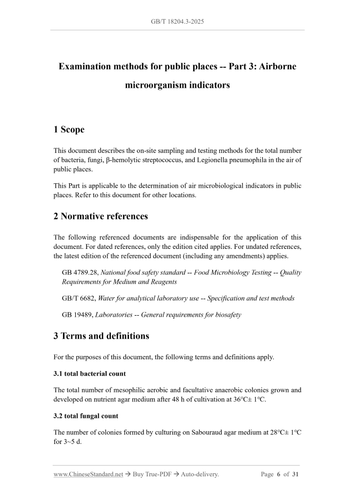
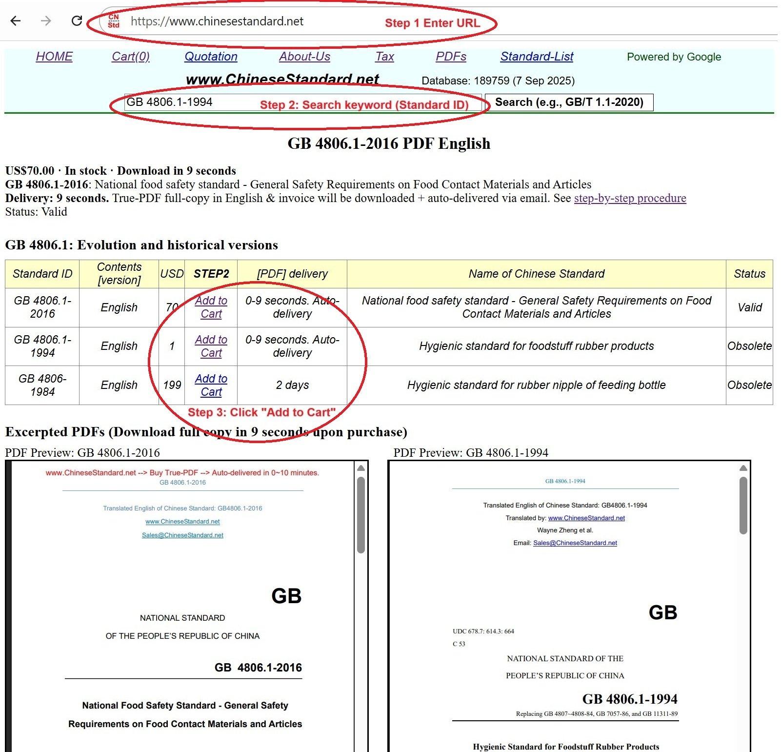
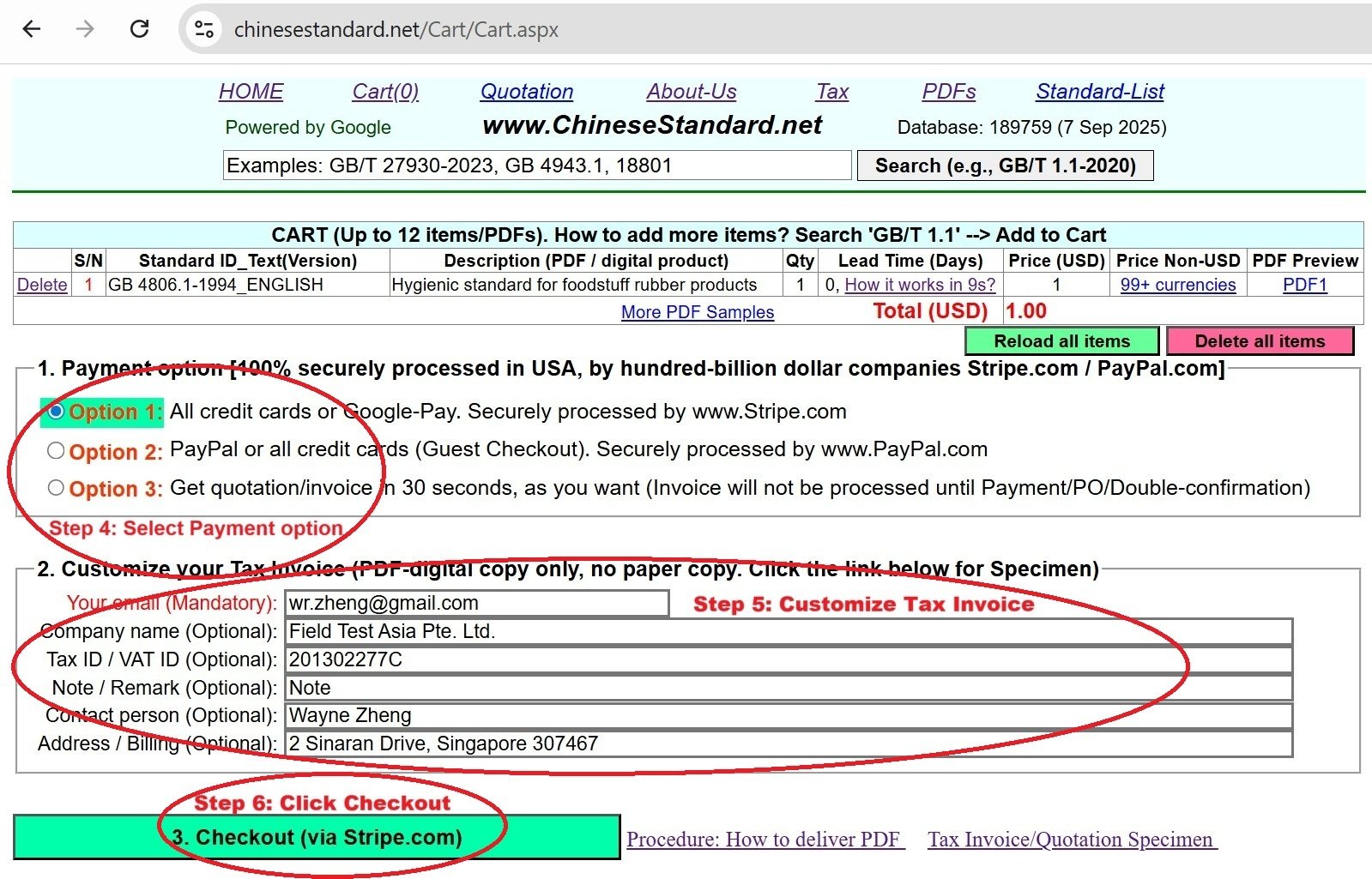
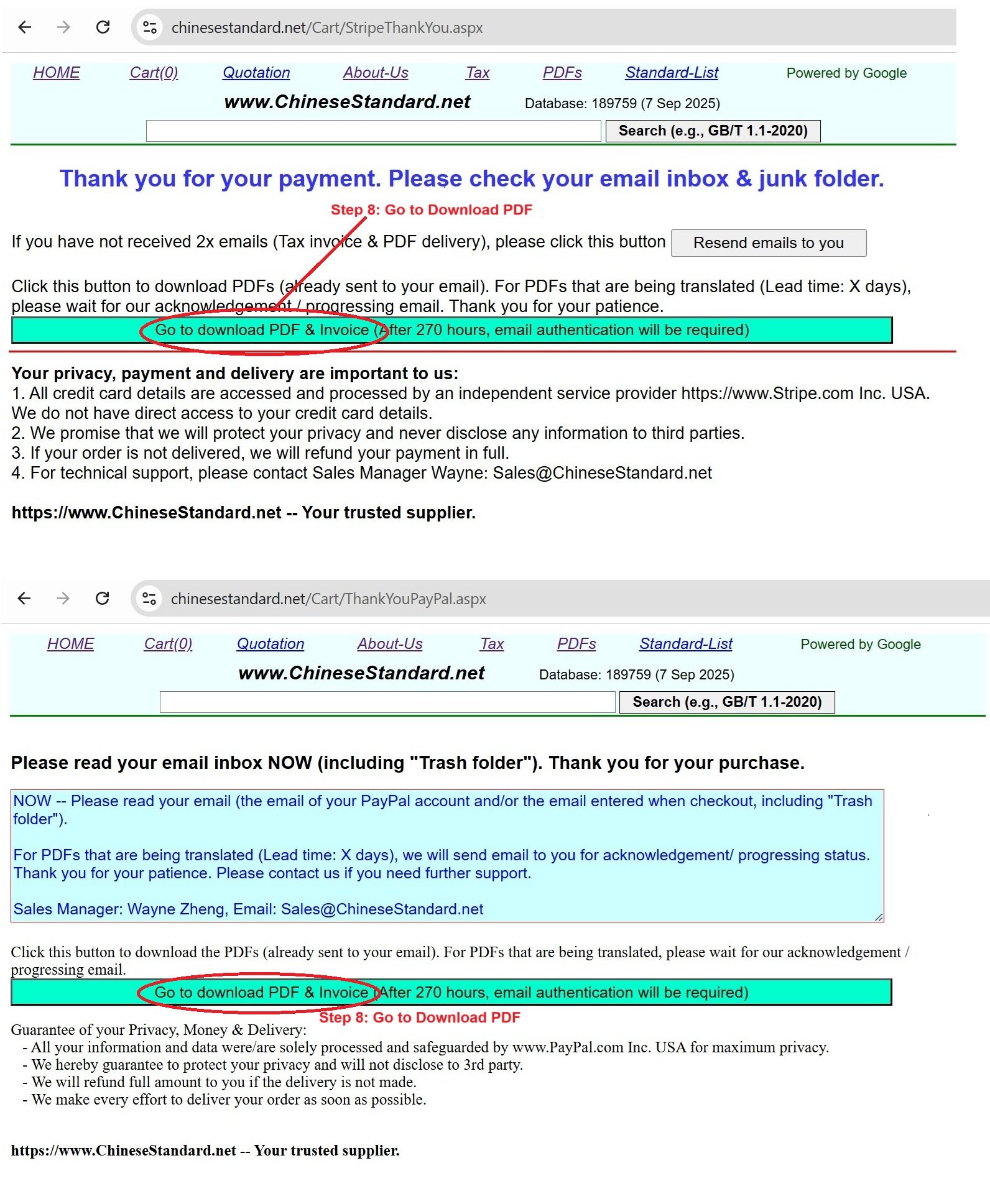
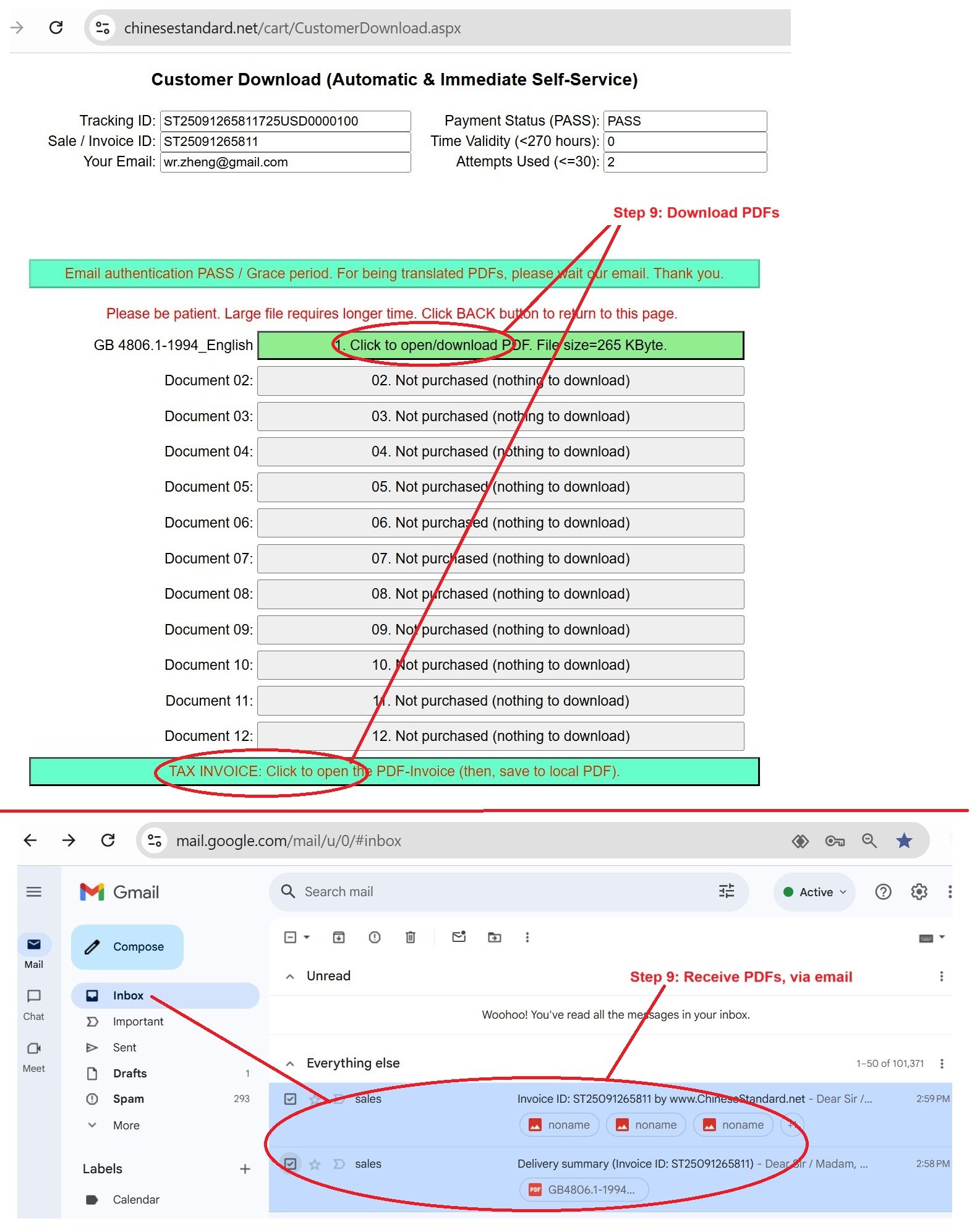
Similar standardsGB/T 18204.3-2025: Examination methods for public places - Part 3: Airborne microorganism indicators---This is an excerpt. Full copy of true-PDF in English version (including equations, symbols, images, flow-chart, tables, and figures etc.), auto-downloaded/delivered in 9 seconds, can be purchased online: https://www.ChineseStandard.net/PDF.aspx/GBT18204.3-2025 GB NATIONAL STANDARD OF THE PEOPLE’S REPUBLIC OF CHINA ICS 13.060 CCS C 51 Replacing GB/T 18204.3-2013 Examination methods for public places -- Part 3.Airborne microorganism indicators Issued on: MAY 30, 2025 Implemented on: DECEMBER 01, 2025 Issued by. State Administration for Market Regulation; Standardization Administration of the People's Republic of China. Table of ContentsForeword... 3 Introduction... 5 1 Scope... 6 2 Normative references... 6 3 Terms and definitions... 6 4 Total bacterial count... 7 5 Total fungal count... 11 6 β-hemolytic streptococcus... 14 7 Legionella pneumophila... 18 Annex A (normative) Requirements for on-site sampling and testing points... 29 Bibliography... 31 Examination methods for public places -- Part 3.Airborne microorganism indicators1 ScopeThis document describes the on-site sampling and testing methods for the total number of bacteria, fungi, β-hemolytic streptococcus, and Legionella pneumophila in the air of public places. This Part is applicable to the determination of air microbiological indicators in public places. Refer to this document for other locations.2 Normative referencesThe following referenced documents are indispensable for the application of this document. For dated references, only the edition cited applies. For undated references, the latest edition of the referenced document (including any amendments) applies. GB 4789.28, National food safety standard -- Food Microbiology Testing -- Quality Requirements for Medium and Reagents GB/T 6682, Water for analytical laboratory use -- Specification and test methods GB 19489, Laboratories -- General requirements for biosafety3 Terms and definitionsFor the purposes of this document, the following terms and definitions apply. 3.1 total bacterial count The total number of mesophilic aerobic and facultative anaerobic colonies grown and developed on nutrient agar medium after 48 h of cultivation at 36℃± 1℃. 3.2 total fungal count The number of colonies formed by culturing on Sabouraud agar medium at 28℃± 1℃ for 3~5 d. 3.3 β-hemolytic streptococcus Streptococcus pyogenes (or Group A) and Streptococcus agalactiae (or Group B) that can produce hemolysin and form clearly defined and completely transparent hemolytic rings (β - type hemolysis) around bacterial colonies on a blood plate. [Source. GB 4789.11-2014,2.2, modified] 3.4 Legionella pneumophila A Gram-negative bacterium with blunt ends, flagella, no spores or capsules, characterized by growth on an activated carbon yeast extract medium containing L- cysteine and trivalent iron salt buffer. It has been identified as a pathogenic Legionella bacterium through biochemical and serological tests and is the main pathogen causing Legionellosis. [Source. GB/T 40392-2021, 3.2, modified] 3.5 impacting method A sampling method that uses an impact type air microbial sampler to collect microorganisms suspended in the air onto the sampling medium through inertial impact. 3.6 natural sinking method A sampling method that involves exposing the culture medium plate to air and allowing microorganisms to naturally settle onto the plate by gravity.4 Total bacterial count4.1 Impact method 4.1.1 Principle Use an impact type air microbial sampler. Generate high-speed airflow by passing air through narrow slits or small holes. Collect microorganisms suspended in the air onto nutrient agar plates. The bacterial colony count was obtained after 48 h of cultivation at 36℃± 1℃. 4.1.2 Instruments and consumables The main instruments and consumables used in this method are as follows. - Six level sieve impact microbial sampler; - High pressure steam sterilizer; - Constant temperature incubator. 36℃± 1℃; - Aseptic petri dish. diameter of 90 mm; - pH meter or precision pH test paper. 4.1.3 Nutrient agar medium 4.1.3.1 Composition Protein peptone. 10.0 g. Beef extract. 3.0 g. Sodium chloride. 5.0 g. Agar. 20.0 g. Pure water. 1000 mL. Finished culture medium that meets the requirements of GB 4789.28 can also be used. The quality of pure water should meet the requirements of Grade Three water or above in GB/T 6682. 4.1.3.2 Preparation method Dissolve peptone, beef extract, and sodium chloride in pure water. Correct the pH to 7.4 ± 0.2.Add agar. Perform high-pressure sterilization at 121℃ for 15 min. When cooled to 45℃~50℃, pour 15mL of culture into each dish based on sterile operation. After cooling, make it into a flat plate for later use. 4.1.4 Sampling 4.1.4.1 Sampling points. shall comply with the requirements of A.1 in Annex A. 4.1.4.2 Sampling environmental conditions. Close doors and windows for 15~30 min during sampling. When sampling in public places where it is difficult to close doors and windows (such as shopping malls, waiting rooms, etc.), the environmental conditions at that time should be maintained. Record the operation status of indoor air conditioning and other equipment, door and window conditions, number of personnel, temperature and humidity, and weather conditions. 4.1.4.3 Sampling method. Aseptic operation should be performed. Load the nutrient agar plates step by step into a six-stage sieve impact microbial sampler. Collect data at a flow rate of 28.3 L/min for 5~15 min. The sampler should be used according to the instructions. 4.1.4.4 Storage and transportation of samples. Store the collected tablets in an environment of 4℃. Return to the laboratory for cultivation within 4 h. 4.1.5 Inspection steps Invert the collected nutrient agar plates and incubate them in a 36℃± 1℃ incubator for 48 h ± 2 h. Perform colony counting. 4.1.6 Inspection results 4.1.6.1 Result calculation The total bacterial concentration in the air of public places is calculated according to They shall meet the requirements of 4.1.5. 4.2.6 Inspection results 4.2.6.1 Result calculation Count the number of bacterial colonies growing on each plate. 4.2.6.2 Result report The measurement results of the total number of bacteria in the air of a region are reported as the average of the total number of bacteria measured at all sampling points in the region. The test results are expressed in colony forming units per plate (CFU/plate). The calculated total number of bacteria should be rounded to the nearest integer. 4.2.7 Quality control It should meet the requirements of 4.1.7.1~4.1.7.5.5 Total fungal count5.1 Impact method 5.1.1 Principle Use an impact type air microbial sampler. Generate high-speed airflow through narrow slits or small holes to collect microorganisms suspended in the air onto a Sabouraud agar plate. The fungal colony count was obtained after culturing at 28℃±1℃ for 3~5 d. 5.1.2 Instruments and consumables The instruments and consumables used in this method are as follows. - Six level sieve impact microbial sampler; - High pressure steam sterilizer; - Fungal incubator. 28℃± 1℃; - Aseptic petri dish. diameter of 90 mm; - pH meter or precision pH test paper. 5.1.3 Sabouraud agar medium 5.1.3.1 Composition Ni - The number of colonies per level of plate, in colony forming units (CFU); v - Sampling flow rate, in liters per minute (L/min); t - Sampling time, in minutes (min); i - Number of tablets. 5.1.6.2 Result report The determination of total fungal count in the air of a region is reported based on the maximum value of total fungal count measured at all sampling points in that region. The test results are expressed in colony forming units per cubic meter (CFU/m3). 5.1.7 Quality control 5.1.7.1 After each batch of culture medium plates is configured, a sterile test should be conducted. Three tablets can be selected per batch. Cultivate and observe according to the steps in 5.1.5.The result should be free of bacterial growth. 5.1.7.2 Other quality controls shall comply with the requirements of 4.1.7.2~4.1.7.5. 5.2 Natural sinking method 5.2.1 Principle Expose the Sabouraud agar plate to air. Microorganisms rely on gravity to naturally settle onto a flat surface. After laboratory cultivation, the total fungal count shall be obtained. 5.2.2 Instruments and consumables The instruments and consumables used in this method are as follows. - High pressure steam sterilizer; - Fungal incubator. 28℃± 1℃; - Sampling bracket; - Aseptic petri dish. diameter of 90mm; - pH meter or precision pH test paper. 5.2.3 Sabouraud agar medium It shall meet the requirements of 5.1.3. 5.2.4 Sampling 5.2.4.1 Sampling points. should meet the requirements of A.2. 5.2.4.2 Sampling environmental conditions. shall comply with the requirements of 4.1.4.2. 5.2.4.3 Sampling method. Aseptic operation should be performed. Place the Sabouraud agar plate at the sampling point. Open the lid of the dish. Expose for 5 min and cover the dish with a lid. 5.2.4.4 Sample transportation and storage. shall comply with the requirements of 4.1.4.4. 5.2.5 Inspection steps They should meet the requirements of 5.1.5. 5.2.6 Inspection results 5.2.6.1 Result calculation Count the number of fungal colonies growing on each plate. 5.2.6.2 Result report The measurement results of total fungal count in the air of a region are reported as the average of total fungal count measured at all sampling points in the region. The test results are expressed in colony forming units per plate (CFU/plate). The calculated total fungal count should be rounded to the nearest integer. 5.2.7 Quality control It should comply with the requirements of 5.1.7.1 and 4.1.7.3~4.1.7.5.6 β-hemolytic streptococcus6.1 Principle Use an impact type air microbial sampler. Generate high-speed airflow through narrow slits or small holes to collect microorganisms suspended in the air onto a blood agar plate. After culturing at 36℃± 1℃ for 18~24 h, counting and biochemical identification are performed to obtain the bacterial count of β-hemolytic streptococcus. 6.2 Instruments and consumables The instruments and consumables used in this method are as follows. - Six level sieve impact microbial sampler; 7.1.3.2 BCYE-Cys culture medium The preparation method is the same as BCYE medium plate, except that L-cysteine hydrochloride is not added. 7.1.3.3 GVPC medium NOTE. This culture medium is based on BCYE supplemented with three antibiotics and glycine. 7.1.3.3.1 GVPC additives 0.3 g/L glycine, polymyxin sulfate B 80000 IU/L, 0.001 g/L vancomycin, 0.08 g/L actinomycin. WARNING. Actinobacillone has hepatotoxicity. Gloves and dust masks should be worn when handling powdered chemical products. 7.1.3.3.2 Preparation of GVPC additives Dissolve an appropriate amount of polymyxin B sulfate (usually 200 mg) in 100 mL of pure water to a concentration of 14545 IU/mL. After mixing, perform membrane filtration for sterilization. Use sterile tubes for subpackaging, 5.5 mL per tube. Store at -25℃~-15℃. Use at room temperature before reusing. Dissolve 20 mg of vancomycin hydrochloride in 20 mL of pure water. After mixing, perform membrane filtration for sterilization. Use sterile tubes for subpackaging. Store 1 mL per tube at -25℃~-15℃. Use at room temperature before reusing. Dissolve 2 g of actinomycete ketone in 100 mL of pure water. After mixing, perform membrane filtration for sterilization. Use sterile tubes for subpackaging, 4 mL per tube. Store at -25℃~-15℃. Use at room temperature before reusing. The maximum freezing storage time for the above antibiotic additives is 6 months. 7.1.3.3.3 Preparation of GVPC culture medium Prepare according to the BCYE culture medium preparation method. After adding the single potassium salt of α-ketoglutarate, add 3 g of glycine. Adjust the pH to 6.8 ± 0.2. After adding dissolved L-cysteine and iron pyrophosphate, three additional antibiotic additives are added. Mix well. 7.1.3.4 Gram staining solution It shall meet the requirements of 6.3.2. 7.1.3.5 Malaurate biochemical reaction tube pH should be 6.9 ± 0.1. 7.1.3.9 Sampling absorption solution Weigh 12.0 g of yeast extract and add pure water to 1000 mL. Sterilize under high pressure at 121℃ for 15 min. Subpack in sterilized centrifuge tubes for later use. 7.1.3.10 Oxidase reagents Weigh 1.0 g of tetramethyl p-phenylenediamine hydrochloride. Dissolve it in 100 mL of pure water for later use. 7.1.3.11 Acid buffer solution 7.1.3.11.1 Hydrochloric acid solution [c (HCl)=0.2 mol/L] Add 17.4 mL of concentrated hydrochloric acid to 1000 mL of pure water. 7.1.3.11.2 Potassium chloride solution [c (KCl)=0.2 mol/L] Dissolve 14.9 g of potassium chloride in 1000 mL of pure water. 7.1.3.11.3 Preparation of acid buffer solution Take 3.9 mL of hydrochloric acid solution [c (HCl)=0.2 mol/L] and 25 mL of potassium chloride solution [c (KCl)=0.2 mol/L]. After mixing, adjust with 1 mol/L potassium hydroxide. Measure the final pH to 2.2 ± 0.2 using precision pH test strips or pH meters. Place in a glass bottle with a stopper. The solution should be kept away from light at room temperature for no more than one month. 7.1.3.12 5% (hydrated) indanone solution Dissolve 1.75 g of (hydrated) indanone in a mixture of 25 mL of acetone and 25 mL of butanol. The solution should be refrigerated in the dark for a maximum of 7 days. 7.1.3.13 Diagnosis serum for Legionella pneumophila Commercialized Legionella pneumophila diagnostic serum can be used. 7.1.4 Sampling 7.1.4.1 Sampling points. shall comply with the requirements of A.1. 7.1.4.2 Sampling environmental conditions. shall comply with the requirements of 4.1.4.2. 7.1.4.3 Sampling method. Pour 20 mL of sampling absorption solution into the aerosol sampler. Operate according to the sampler manual. The air volume collected for each aerosol sample is 2 m3.When using the same aerosol sampler to collect air samples a CO2 concentration (volume fraction) of 5%. Cultivate at 36℃± 1℃ for 2 d. If there are different types of suspected Legionella colonies, at least 3 suspected colonies of each type should be selected for culture validation. Legionella colonies grow on BCYE but not on BCYE-Cys plates. Record the results of each tablet. Biochemical identification of Legionella colonies. 7.1.5.6 Biochemical identification 7.1.5.6.1 Bacterial staining. Gram staining microscopy is performed on bacterial colonies. Observe the morphology and color of bacteria. If the bacterial cells are purple, they are Gram positive bacteria. If the bacterial cells are red, they are Gram negative bacteria. 7.1.5.6.2 Oxidase test. Use a platinum/iridium inoculation ring or glass rod to pick up the test colony and place it on filter paper. Add 1 drop of oxidase reagent. If violet or dark blue appears within 30 s, it is considered positive; If there is no color change within 2 min, it is considered negative. 7.1.5.6.3 Nitrate reduction test. Inoculate the test bacteria onto nitrate culture medium. Cultivate at 36℃± 1℃ for 1~3 d. Add 1 drop each of solution A and solution B. Observe the results. If it turns red immediately or within a few minutes, it is considered positive. If there is no change in the color of the culture medium, it is considered negative. 7.1.5.6.4 Urease test. Inoculate a large amount of the test bacteria into urea agar medium through puncture. Cultivate at 36℃± 1℃ for 2 and 24 h. Observe the results separately. If the culture medium turns red, it is considered positive. If the color remains unchanged, it is negative. 7.1.5.6.5 Gelatin liquefaction test. Inoculate the test bacteria into gelatin culture medium by puncture. Cultivate at 36℃± 1℃ for 24 h ± 2 h. Remove and place in a refrigerator at 4℃± 2℃ for 10~30 min before observing the results. When it is still in a dissolved state or on the surface, it is considered positive in the gelatin liquefaction test. Those who are insoluble in coagulation are negative. 7.1.5.6.6 Uric acid hydrolysis test. Select the bacterial colony to be tested. Add it to a test tube containing 0.4 mL of 1% sodium hippurate to prepare a bacterial suspension. After mixing evenly, incubate in a water bath at 36℃± 1℃ or in a 36℃± 1℃ incubator for 24 h. Slowly add 0.2 mL of indanone solution along the wall of the test tube. After incubating in a 36℃± 1℃ water bath or placing in a 36℃± 1℃ incubator for 10 min, observe the results. If the solution shows a blue purple change within 15 min, it is considered positive. If there is no color change in the solution or a blue purple change occurs after 15 min, it is considered negative. In order to avoid false negatives during the test, standard bacterial strains are used as positive controls. 7.1.5.6.7 Biochemical culture result determination. Gram negative non spore forming bacteria detected by staining microscopy, with oxidase (-/weak+), nitrate reduction (-), urease (-), gelatin liquefaction (+), and hydrolyzed hippuric acid (+), can be confirmed k - Suspected type of Legionella pneumophila (i=1,2,3,..., k). 7.1.6.2 Result report Qualitative result report. Any sampling point in a region is positive for Legionella pneumophila, indicating that the measurement result of Legionella pneumophila in the air of the region is positive. Otherwise, report 'No Legionella pneumophila detected'. Quantitative result report. The quantitative detection results of Legionella pneumophila in the air of a region are given based on the maximum value of Legionella pneumophila measured at all sampling points in the region. 7.1.7 Quality control 7.1.7.1 The culture medium and reagents should meet the quality requirements of GB 4789.28.The purchased finished culture medium should be used within its validity period. 7.1.7.2 Before sampling begins, ensure that the reagents and materials used are in a sterile state. Aseptic operation should be carried out during the operation process and sample transportation. Avoid human pollution. 7.1.7.3 A positive control should be set up (Legionella pneumophila standard strain ATCC 33152 or other equivalent standard strains). 7.1.7.4 The sampler should be calibrated regularly under load conditions using a flow meter that has been verified/calibrated to be qualified. The relative deviation should not exceed 5%, or be calibrated according to the instructions of the sampler. Before sampling, the airtightness of the sampling system should be checked to ensure that there is no air leakage. 7.1.7.5 The personal protection of experimental personnel and the disposal of testing waste should be carried out in accordance with GB 19489.As Legionella pneumophila is a pathogenic bacterium, experimental operators should have practical experience in working in a secondary biosafety laboratory. 7.2 Fluorescent polymerase chain (PCR) rapid detection method 7.2.1 Principle Specific primers and probes are designed for the highly conserved region of the mip gene in Legionella pneumophila. In the presence of a Legionella pneumophila genome template in the reaction system, polymerase chain reaction (PCR) is performed and fluorescent signals are released. The instrument is used to monitor and output the signal intensity of the corresponding channel during the PCR process in real time, so as to achieve qualitative analysis of the test results. It is suitable for initial screening of Legionella pneumophila and rapid identification of suspicious bacterial colonies. ......Source: Above contents are excerpted from the full-copy PDF -- translated/reviewed by: www.ChineseStandard.net / Wayne Zheng et al. Tips & Frequently Asked Questions:Question 1: How long will the true-PDF of English version of GB/T 18204.3-2025 be delivered?Answer: The full copy PDF of English version of GB/T 18204.3-2025 can be downloaded in 9 seconds, and it will also be emailed to you in 9 seconds (double mechanisms to ensure the delivery reliably), with PDF-invoice.Question 2: Can I share the purchased PDF of GB/T 18204.3-2025_English with my colleagues?Answer: Yes. The purchased PDF of GB/T 18204.3-2025_English will be deemed to be sold to your employer/organization who actually paid for it, including your colleagues and your employer's intranet.Question 3: Does the price include tax/VAT?Answer: Yes. Our tax invoice, downloaded/delivered in 9 seconds, includes all tax/VAT and complies with 100+ countries' tax regulations (tax exempted in 100+ countries) -- See Avoidance of Double Taxation Agreements (DTAs): List of DTAs signed between Singapore and 100+ countriesQuestion 4: Do you accept my currency other than USD?Answer: Yes. www.ChineseStandard.us -- GB/T 18204.3-2025 -- Click this link and select your country/currency to pay, the exact amount in your currency will be printed on the invoice. Full PDF will also be downloaded/emailed in 9 seconds.Question 5: Should I purchase the latest version GB/T 18204.3-2025?Answer: Yes. Unless special scenarios such as technical constraints or academic study, you should always prioritize to purchase the latest version GB/T 18204.3-2025 even if the enforcement date is in future. Complying with the latest version means that, by default, it also complies with all the earlier versions, technically.How to buy and download a true PDF of English version of GB/T 18204.3-2025?A step-by-step guide to download PDF of GB/T 18204.3-2025_EnglishStep 1: Visit website https://www.ChineseStandard.net (Pay in USD), or https://www.ChineseStandard.us (Pay in any currencies such as Euro, KRW, JPY, AUD).Step 2: Search keyword "GB/T 18204.3-2025". Step 3: Click "Add to Cart". If multiple PDFs are required, repeat steps 2 and 3 to add up to 12 PDFs to cart. Step 4: Select payment option (Via payment agents Stripe or PayPal). Step 5: Customize Tax Invoice -- Fill up your email etc. Step 6: Click "Checkout". Step 7: Make payment by credit card, PayPal, Google Pay etc. After the payment is completed and in 9 seconds, you will receive 2 emails attached with the purchased PDFs and PDF-invoice, respectively. Step 8: Optional -- Go to download PDF. Step 9: Optional -- Click Open/Download PDF to download PDFs and invoice. See screenshots for above steps: Steps 1~3 Steps 4~6 Step 7 Step 8 Step 9 |